menu
Stanford Anatomic Pathology & Clinical Laboratories
Search
  • Anatomic Pathology Anatomic Path...
  • Clinical Pathology Clinical Path...
  • Molecular Pathology/Genomics Molecular Path...
  • Transfusion Medicine/Blood Center Transfusion Medicine/Blood Center
  • Reference Lab Testing Reference Lab Testing
  • Test Directory Tests
  • Autopsy Pathology
  • Cytopathology
  • Dermatopathology
  • Hematopathology
  • Neuropathology
  • Ophthalmic Pathology
  • Pediatric Pathology
  • Surgical Pathology
  • Breast
  • Cardiovascular
  • Gastrointestinal
  • Genitourinary
  • Gynecologic
  • Head & Neck
  • Liver
  • Pediatric & Perinatal
  • Pulmonary & Thoracic
  • Renal
  • Soft Tissue & Bone
  • Consulting Services
  • Ancillary Testing
  • Immunohistochemistry
  • Cytogenetics/FISH
  • Electron Microscopy
  • Immunofluorescence
  • Clinical Genomics
  • Molecular Pathology
  • Flow Cytometry


  • View All Info
  • Biochemical Genetics
  • Clinical Chemistry
  • Flow Cytometry
  • Hematology
  • Hematopathology
  • Histocompatibility and Immunogenetics Lab (HLA)
  • Immunology & Special Chemistry
  • Microbiology
  • Special Coagulation
  • Virology
  • COVID-19 Resource Center


  • View All Info
  • Biochemical Genetics
  • Molecular Genetic Pathology
  • Cytogenetics/FISH
  • Clinical Genomics
  • View All Info
  • Transfusion Medicine
  • Histocompatibility and Immunogenetics Lab (HLA)
  • Stanford Blood Center
  • View All Info
Home
Close
Anatomic Pathology
Clinical Pathology
Molecular Pathology/Genomics
Transfusion Medicine/Blood Center
Reference Lab Testing
Test Directory

Submit a Case/Specimen

Contact Us
About Us Careers Billing
Patient Information
Clear

Electron Microscopy

A Diagnostic Modality Supporting  Anatomic Pathology

Stanford’s Electron Microscopy Lab

Electron microscopy (EM) is a technique for obtaining high resolution images of specimens. It is used in the evaluation of pathology cases for detailed examination of structure of tissues, cells, viruses, organelles and macromolecular complexes. The high resolution of EM images results from using a beam of accelerated electrons as the source of illuminating radiation. These images provide key information on the structural basis of cell function and of cell disease.


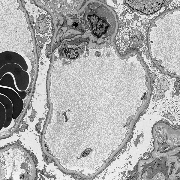
Stanford’s Electron Microscopy Service primarily assists in the diagnosis of medical kidney disease, heart and skeletal muscle disorders, neurological disorders, viral infections and ciliary dysfunction. Our team of highly experienced and specialized Electron Microscopy Technologists bring decades of experience to the processing of EM cases, ensuring that cases are efficiently and expertly handled.

Contact Information

Immunofluorescence/Electron Microscopy Lab

650-725-5196

Monday-Friday 8 a.m. to 5 p.m.


Related Collection Guides:

  • Renal Biopsy Collection Guide
  • Epidermolysis Bullosa Collection Guide
  • Ciliary Dyskinesia Collection Guide
 
Medical Director:
Megan Troxell, MD, PhD

Megan Troxell, MD, PhD

More information about Megan Troxell

  • PATHOLOGY CONSULTATIONS & REFERENCE LAB TESTING
  • Requisitions
  • Client Logistics
  • Billing
  • Program FAQ’s
  • Web Results Portal
  • ABOUT US
  • Contact Us
  • Licensure
  • NEWS
  • CAREERS
  • FIND A TEST
  • PATIENT INFO
  • Stanford Medicine
  • Stanford Health Care
Stanford Medicine
Website Terms of Use & Privacy Policy |
© 2025 Stanford Health Care. All Rights Reserved.
Close