
Dermatopathology
A Subspecialty Of Anatomic Pathology
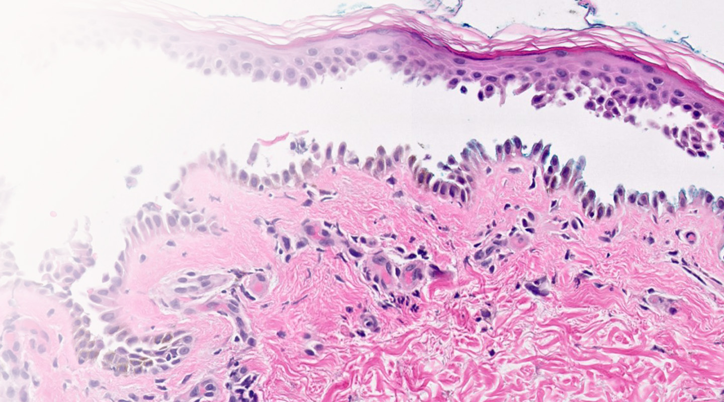
About Dermatopathology
Dermatopathology is a subspecialty of Pathology that provides diagnosis on biopsies from the skin, mucous membranes, and adjacent soft tissues to identify skin cancer and a range of other skin conditions.
Our Services
Submit a Specimen
Our Faculty
Ancillary Testing
Stanford’s Dermatopathology Service reviews cases for both primary diagnosis and consultation/second opinion by board certified pathologists and dermatopathologists. Our wide spectrum of services includes routine sectioning, special histochemistry and immunohistochemistry, immunofluorescence, and horizontal sections for evaluation of alopecia.
Primary Pathology Diagnosis and Consultative Case Reviews
Stanford’s Dermatopathology Service reads the full range of wet tissue and slide consult cases. In addition to the expert diagnosis provided by our Dermatopathologists, challenging cases are often shared among other faculty members in Surgical Pathology, Soft Tissue Pathology, and Hematopathology, ensuring that the patient’s diagnosis is made with consideration of a team of experts.
Biopsies for Alopecia
Stanford Dermatopathology provides interpretation of scalp biopsies for alopecia. Such biopsies are embedded horizontally and examined according to an alopecia protocol. The diagnostic yield of a horizontally embedded scalp biopsy is far superior that gleaned from a vertically embedded specimen. When submitting a biopsy to assess the type of alopecia, please check the appropriate box on our requisition or write the word “alopecia” in a prominent position on the page.
Epidermolysis Bullosa
Stanford Dermatopathology evaluates biopsies for the diagnosis of epidermolysis bullosa (EB) utilizing electron microscopy and utilizing immunomapping of the basement membrane zone.
We provide this service in close collaboration with Stanford’s Pediatric Dermatology service at Stanford and our Clinic for Bullous Disorders, ensuring that a team of experts in this subject are reviewing the case.
Our antibody panel for mapping of the basement membrane zone includes:
mA6K5, anti-keratin 5
mAb K14, anti-keratin 14
mAb HD121, anti-plectin
mAb 123, collagen XVII (BP180)
mAb 3E1, anti-beta 4 integrin
mAb K140, anti-laminin-332 beta 3 chain
mAb D4B5, anti-laminin-332 gamma 2 chain
mAb BM165, anti-laminin-332 alpha 3 chain
mAb AE2, anti-type IV collagen
mAb LH-72, anti-type VII collagen
Primary panel: Antibodies against collagen XVII (BP180), collagen IV, collagen VII, laminin-332 gamma 2 chain
Secondary panel: Antibodies against laminin-332 beta 3 chain, laminin-332 alpha 3 chain, and beta 4 integrin
Antibodies against keratin 5, keratin 14, and plectin are run on an as-needed basis.
Specimen collection for EB requires special handling. Please see our collection guide for more information.
Consult Cases
To submit slides for case review, please complete a Stanford requisition and include all slides, relevant reports, and ancillary testing materials and reports. If possible, please include a block, as it will greatly speed the finalization of the case should additional testing be necessary. Package all materials securely and send via a padded envelope.
Though not necessary, we strongly encourage the submission of clinical photographs of patients who undergo biopsy for a rash or who present with unusual pigmented or non-pigmented neoplasms. Electronic submission of digital photographs is simple via our secure email address. This e-mail account is secure and can only be accessed by faculty, fellows and staff in Dermatopathology. Please maintain patient privacy and do not include full facial photographs or patient names in the e-mail.
E-mail: secure-dermpath@lists.stanford.edu
Wet Tissue Specimens
Please submit materials in proper transport media. All samples should be labeled with the patient information (name, DOB and one other unique identifier) and sealed in a biohazard bag. Please submit a signed Stanford requisition with each sample.
Epidermolysis Bullosa
Cases submitted for evaluation of epidermolysis bullosa (EB) require special handling and collection of the specimen.
Samples and consult materials should be shipped or delivered via courier to:
Stanford Dermatopathology
300 Pasteur Drive, Room H2110
Stanford, CA 94305
New Client Inquiries
To arrange to be set up as a new client, please contact:
ClinOpsContracts@stanfordhealthcare.org.
Contact Us
To submit a specimen to Dermatopathology, please complete a requisition and send with sample to:
Stanford Dermatopathology
300 Pasteur Drive, Room H2110
Stanford, CA 94305
Email: secure-dermpath@lists.stanford.edu
Medical Director:
Kerri Rieger, MD
To arrange to be set up as a new client, please contact:
ClinOpsContracts@
stanfordhealthcare.org
SAMPLE SUBMISSION LOGISTICS
For more information on submitting cases, see the links below:
All Requisitions
Download Stanford Requisitions for specimen submission.
Account Set-up
Information on personalized requisitions, supplies, shipping and courier pick-up.
Pricing Inquiries
Pricing inquiries and contract information.
Billing
Billing policies, information and questions.
Results Web Portal
Request access or log-in to our client results portal.
Specimen Collection and Handling
Specifications for Specimen Collection and Handling.
Consult Service FAQ's
Get answers to Frequently Asked Questions.
Kerri Rieger, MD, PhD

APPOINTMENTS
- Clinical Associate Professor of Pathology (Dermatopathology) and Dermatology
- Director of Dermatopathology Service, Director of Dermatopathology Fellowship Program
PRACTICE AREAS
- Adult General Dermatology
- Dermatopathology
CONTACT
Email: kerieger@stanford.edu
Phone: (650) 725-9860
More information about Kerri Rieger:
https://med.stanford.edu/profiles/kerri-rieger
https://profiles.stanford.edu/kerri-rieger
Roberto Novoa, MD

APPOINTMENTS
- Clinical Assistant Professor of Pathology (Dermatopathology) and Dermatology
- Associate Director of Dermatopathology Fellowship Program
PRACTICE AREAS
- Dermatology
- Dermatopathology
CONTACT
Email: rnovoa@stanford.edu
Phone: (650) 723-6316
More information about Roberto Novoa:
https://med.stanford.edu/profiles/roberto-novoa
https://profiles.stanford.edu/roberto-novoa
Ryanne Brown, MD, MBA

APPOINTMENTS
- Clinical Assistant Professor of Pathology (Dermatopathology)
PRACTICE AREAS
- Anatomic and Clinical Pathology
CONTACT
Email: rbrown85@stanford.edu
Phone: (650) 725-7407
More information about Ryanne Brown:
https://med.stanford.edu/profiles/ryanne-brown
https://profiles.stanford.edu/ryanne-brown
Ancillary Testing
In our interpretation of skin biopsy specimens Stanford Dermatopathology utilizes ancillary techniques as necessary. We practice judicious use of special techniques and do not apply them indiscriminately.
Immunofluorescence »
We perform direct immunofluorescence for autoimmune and blistering disorders on tissue samples and indirect immunofluorescence on serum. We also perform immunomapping of the basement membrane region for epidermolysis bullosa congenita.
Please submit tissue for direct immunofluorescence in Zeus or Michaels transport medium. Once in transport medium, tissue is stable for several weeks at room temperature and can be picked up or shipped with your routine formalin fixed specimens.
Please submit serum for indirect immunofluorescence in a red top tube. We use monkey esophagus as substrate for our indirect assay.
Immunohistochemistry »
Our immunohistochemical laboratory is fully equipped to perform a wide array of antibody stains.
Molecular Pathology »
We perform gene rearrangement studies for T- and B-cell clonality by PCR, tests for microsattelite instability and PCR or FISH for chromosomal translocations. To submit directly to the molecular laboratory, please download the Molecular Genetic Requisition Form (.pdf).
We can also test for microsatellite instability for the Muir Torre or Lynch syndromes via PCR analysis (as an adjunct to immunohistochemical studies).
Fluorescence in situ hybridization (FISH) studies for chromosomal translocations and rearrangements that are associated with particular types of leukemias/lymphomas and solid tumors are performed in the Cytogenetics Laboratory.
Electron Microscopy »
Stanford Dermatopathology utilizes electron microscopy in the evaluation of biopsies for epidermolysis bullosa (EB).
Stanford’s Dermatopathology Service reviews cases for both primary diagnosis and consultation/second opinion by board certified pathologists and dermatopathologists. Our wide spectrum of services includes routine sectioning, special histochemistry and immunohistochemistry, immunofluorescence, and horizontal sections for evaluation of alopecia.
Primary Pathology Diagnosis and Consultative Case Reviews
Stanford’s Dermatopathology Service reads the full range of wet tissue and slide consult cases. In addition to the expert diagnosis provided by our Dermatopathologists, challenging cases are often shared among other faculty members in Surgical Pathology, Soft Tissue Pathology, and Hematopathology, ensuring that the patient’s diagnosis is made with consideration of a team of experts.
Biopsies for Alopecia
Stanford Dermatopathology provides interpretation of scalp biopsies for alopecia. Such biopsies are embedded horizontally and examined according to an alopecia protocol. The diagnostic yield of a horizontally embedded scalp biopsy is far superior that gleaned from a vertically embedded specimen. When submitting a biopsy to assess the type of alopecia, please check the appropriate box on our requisition or write the word “alopecia” in a prominent position on the page.
Epidermolysis Bullosa
Stanford Dermatopathology evaluates biopsies for the diagnosis of epidermolysis bullosa (EB) utilizing electron microscopy and utilizing immunomapping of the basement membrane zone.
We provide this service in close collaboration with Stanford’s Pediatric Dermatology service at Stanford and our Clinic for Bullous Disorders, ensuring that a team of experts in this subject are reviewing the case.
Our antibody panel for mapping of the basement membrane zone includes:
mA6K5, anti-keratin 5
mAb K14, anti-keratin 14
mAb HD121, anti-plectin
mAb 123, collagen XVII (BP180)
mAb 3E1, anti-beta 4 integrin
mAb K140, anti-laminin-332 beta 3 chain
mAb D4B5, anti-laminin-332 gamma 2 chain
mAb BM165, anti-laminin-332 alpha 3 chain
mAb AE2, anti-type IV collagen
mAb LH-72, anti-type VII collagen
Primary panel: Antibodies against collagen XVII (BP180), collagen IV, collagen VII, laminin-332 gamma 2 chain
Secondary panel: Antibodies against laminin-332 beta 3 chain, laminin-332 alpha 3 chain, and beta 4 integrin
Antibodies against keratin 5, keratin 14, and plectin are run on an as-needed basis.
Specimen collection for EB requires special handling. Please see our collection guide for more information.
close Our Services
Consult Cases
To submit slides for case review, please complete a Stanford requisition and include all slides, relevant reports, and ancillary testing materials and reports. If possible, please include a block, as it will greatly speed the finalization of the case should additional testing be necessary. Package all materials securely and send via a padded envelope.
Though not necessary, we strongly encourage the submission of clinical photographs of patients who undergo biopsy for a rash or who present with unusual pigmented or non-pigmented neoplasms. Electronic submission of digital photographs is simple via our secure email address. This e-mail account is secure and can only be accessed by faculty, fellows and staff in Dermatopathology. Please maintain patient privacy and do not include full facial photographs or patient names in the e-mail.
E-mail: secure-dermpath@lists.stanford.edu
Wet Tissue Specimens
Please submit materials in proper transport media. All samples should be labeled with the patient information (name, DOB and one other unique identifier) and sealed in a biohazard bag. Please submit a signed Stanford requisition with each sample.
Epidermolysis Bullosa
Cases submitted for evaluation of epidermolysis bullosa (EB) require special handling and collection of the specimen.
Samples and consult materials should be shipped or delivered via courier to:
Stanford Dermatopathology
300 Pasteur Drive, Room H2110
Stanford, CA 94305
New Client Inquiries
To arrange to be set up as a new client, please contact:
ClinOpsContracts@stanfordhealthcare.org.
Contact Us
To submit a specimen to Dermatopathology, please complete a requisition and send with sample to:
Stanford Dermatopathology
300 Pasteur Drive, Room H2110
Stanford, CA 94305
Email: secure-dermpath@lists.stanford.edu
Medical Director:
Kerri Rieger, MD
To arrange to be set up as a new client, please contact:
ClinOpsContracts@
stanfordhealthcare.org
SAMPLE SUBMISSION LOGISTICS
For more information on submitting cases, see the links below:
All Requisitions
Download Stanford Requisitions for specimen submission.
Account Set-up
Information on personalized requisitions, supplies, shipping and courier pick-up.
Pricing Inquiries
Pricing inquiries and contract information.
Billing
Billing policies, information and questions.
Results Web Portal
Request access or log-in to our client results portal.
Specimen Collection and Handling
Specifications for Specimen Collection and Handling.
Consult Service FAQ's
Get answers to Frequently Asked Questions.
close Submit a Specimen
Kerri Rieger, MD, PhD
APPOINTMENTS
- Clinical Associate Professor of Pathology (Dermatopathology) and Dermatology
- Director of Dermatopathology Service, Director of Dermatopathology Fellowship Program
PRACTICE AREAS
- Adult General Dermatology
- Dermatopathology
CONTACT
Email: kerieger@stanford.edu
Phone: (650) 725-9860
More information about Kerri Rieger:
https://med.stanford.edu/profiles/kerri-rieger
https://profiles.stanford.edu/kerri-rieger
Roberto Novoa, MD
APPOINTMENTS
- Clinical Assistant Professor of Pathology (Dermatopathology) and Dermatology
- Associate Director of Dermatopathology Fellowship Program
PRACTICE AREAS
- Dermatology
- Dermatopathology
CONTACT
Email: rnovoa@stanford.edu
Phone: (650) 723-6316
More information about Roberto Novoa:
https://med.stanford.edu/profiles/roberto-novoa
https://profiles.stanford.edu/roberto-novoa
Ryanne Brown, MD, MBA
APPOINTMENTS
- Clinical Assistant Professor of Pathology (Dermatopathology)
PRACTICE AREAS
- Anatomic and Clinical Pathology
CONTACT
Email: rbrown85@stanford.edu
Phone: (650) 725-7407
More information about Ryanne Brown:
https://med.stanford.edu/profiles/ryanne-brown
https://profiles.stanford.edu/ryanne-brown
close Our Faculty
Ancillary Testing
In our interpretation of skin biopsy specimens Stanford Dermatopathology utilizes ancillary techniques as necessary. We practice judicious use of special techniques and do not apply them indiscriminately.
Immunofluorescence »
We perform direct immunofluorescence for autoimmune and blistering disorders on tissue samples and indirect immunofluorescence on serum. We also perform immunomapping of the basement membrane region for epidermolysis bullosa congenita.
Please submit tissue for direct immunofluorescence in Zeus or Michaels transport medium. Once in transport medium, tissue is stable for several weeks at room temperature and can be picked up or shipped with your routine formalin fixed specimens.
Please submit serum for indirect immunofluorescence in a red top tube. We use monkey esophagus as substrate for our indirect assay.
Immunohistochemistry »
Our immunohistochemical laboratory is fully equipped to perform a wide array of antibody stains.
Molecular Pathology »
We perform gene rearrangement studies for T- and B-cell clonality by PCR, tests for microsattelite instability and PCR or FISH for chromosomal translocations. To submit directly to the molecular laboratory, please download the Molecular Genetic Requisition Form (.pdf).
We can also test for microsatellite instability for the Muir Torre or Lynch syndromes via PCR analysis (as an adjunct to immunohistochemical studies).
Fluorescence in situ hybridization (FISH) studies for chromosomal translocations and rearrangements that are associated with particular types of leukemias/lymphomas and solid tumors are performed in the Cytogenetics Laboratory.
Electron Microscopy »
Stanford Dermatopathology utilizes electron microscopy in the evaluation of biopsies for epidermolysis bullosa (EB).
close Ancillary Testing
Residencies and Fellowships
Anatomic Pathology Residency Programs »
Stanford Pathology offers a 3-year Anatomic Pathology residency and 4 year combination residencies in Anatomic Pathology/Clinical Pathology and Anatomic Pathology/Neuropathology.
Dermatopathology Fellowships »
Stanford offers an ACGME-accredited Fellowship in Dermatopathology.